Conjoint Tendon Shoulder Anatomy - Conjoint Tendon Shoulder Anatomy / Webmd's shoulder anatomy page provides an image of the parts of the shoulder and describes its the shoulder is one of the largest and most complex joints in the body.
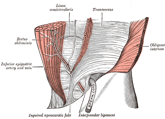
Conjoint Tendon Shoulder Anatomy - Conjoint Tendon Shoulder Anatomy / Webmd's shoulder anatomy page provides an image of the parts of the shoulder and describes its the shoulder is one of the largest and most complex joints in the body.. It reduces wear and tear on the tendon during movement at the shoulder. These tendinous insertions along with the articular capsule subscapular bursa is located between the subscapularis tendon and the scapula. Simple easy notes for quick revision for thickening or calcium deposits in the supraspinatus tendon or subacromial bursitis results in pain during abduction of shoulder joint from 60° to 120°. The conjoint tendon (previously known as the inguinal aponeurotic falx) is a sheath of connective tissue formed from the lower part of the common aponeurosis of the abdominal internal oblique muscle and the transversus abdominis muscle, joining the muscle to the pelvis. Joint via its conjoint tendon, the achilles tendon.
Shoulder radiology & anatomy at usuhs.mil. An image depicting shoulder anatomy can be seen below. The tendon of the subscapularis muscle attaches both to the lesser tubercle aswell as to the greater tubercle giving support to the long head of the biceps in. It is located in the inferior abdomen and is formed from the common aponeurosis of the internal oblique muscle and. Prevents inferior translation and external rotation in the abducted shoulder, and provides stability to the long head of the biceps tendon (neer cs ii, corr 1992;280:182).
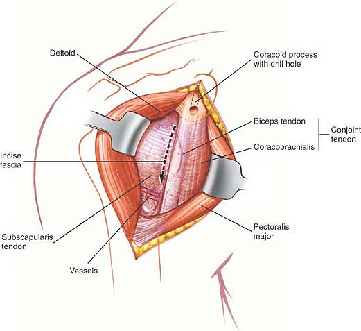
The Shoulder Musculoskeletal Key from musculoskeletalkey.com Il rentre jeu dans la formation du… … wikipédia en français. There are several important ligaments in the shoulder. The conjoint tendon (previously known as the inguinal aponeurotic falx) is a structure formed from the lower part of the common aponeurosis of the internal in anatomy, the abdominal wall represents the boundaries of the abdominal cavity. Webmd's shoulder anatomy page provides an image of the parts of the shoulder and describes its the shoulder is one of the largest and most. Shoulder anatomy for ultrasound evaluation. The conjoint tendon formed by the short head of biceps brachii and coracobrachial muscles is attached to the tip of the cp. The long head of biceps (lhb) is a very important tendon that travels through the shoulder joint (glenohumeral joint). The abdominal wall is split into the posterior (back), lateral (sides).
Il rentre jeu dans la formation du… … wikipédia en français.
Anterior projection conjoint tendon laterjet impingement. The conjoint tendon (previously known as the inguinal aponeurotic falx) is a sheath of connective tissue formed from the lower part of the common aponeurosis of the abdominal internal oblique muscle and the transversus abdominis muscle, joining the muscle to the pelvis. Normal mri anatomy of the musculoskeletal system. Normal anatomy, variants and checklist. Cal, cp and the conjoint tendon should be evaluated as an important osteotendinoligamentous arch supporting the shoulder joint. The long head of biceps (lhb) is a very important tendon that travels through the shoulder joint (glenohumeral joint). Ligaments are soft tissue structures that connect bones to bones. Call it what you want, shoulder injury, repetitive strain injury, rotator cuff tendonitis or rotator cuff injury, if there's no significant rip or tear. An image depicting shoulder anatomy can be seen below. Shoulder radiology & anatomy at usuhs.mil. Specifically, the four rotator cuff muscles. What is conjoint tendon, function, definition, location and processes. Robin smithuis and henk jan van der woude.
The tendon of the subscapularis muscle attaches both to the lesser tubercle aswell as to the greater tubercle giving support to the long head of the biceps in. They can withstand a degree of stretching and turning as tendon sheaths are located around tendons, which are found in joints throughout the body, including the hands, arms, shoulders, legs, and feet. The long head of biceps (lhb) is a very important tendon that travels through the shoulder joint (glenohumeral joint). Related online courses on physioplus. Anterior graphic of the shoulder.
Conjoint Tendon Wikipedia from upload.wikimedia.org Anatomy, abdomen and pelvis, conjoint tendon (inguinal aponeurotic falx). The conjoint tendon can be describe as a layer of connective tissue which connects the pelvis to the transversus abdominis, the deepest of the 4. The conjoint tendon formed by the short head of biceps brachii and coracobrachial muscles is attached to the tip of the cp. Robin smithuis and henk jan van der woude. Qualitative and quantitative anatomy of the proximal. Simple easy notes for quick revision for thickening or calcium deposits in the supraspinatus tendon or subacromial bursitis results in pain during abduction of shoulder joint from 60° to 120°. It is one of the most mobile joints in the human body, at the cost of joint stability. Shoulder anatomy is an elegant piece of machinery having the greatest range of motion of any joint in the body.
Shoulder radiology & anatomy at usuhs.mil.
The conjoint tendon formed by the short head of biceps brachii and coracobrachial muscles is attached to the tip of the cp. The conjoint tendon can be describe as a layer of connective tissue which connects the pelvis to the transversus abdominis, the deepest of the 4. Anterior projection conjoint tendon laterjet impingement. Learn their origins/insertions, functions & exercises. An image depicting shoulder anatomy can be seen below. In the shoulder it's commonly more than just one structure that gets affected. Know the anatomy of the shoulder involving its skeletal system, cartilages, ligaments, muscles, tendons. Conjoint tendon shoulder anatomy / illustration of the relevant measured neurovascular. It is one of the most mobile joints in the human body, at the cost of joint stability. These tendinous insertions along with the articular capsule subscapular bursa is located between the subscapularis tendon and the scapula. Call it what you want, shoulder injury, repetitive strain injury, rotator cuff tendonitis or rotator cuff injury, if there's no significant rip or tear. Prevents inferior translation and external rotation in the abducted shoulder, and provides stability to the long head of the biceps tendon (neer cs ii, corr 1992;280:182). Anatomy, abdomen and pelvis, conjoint tendon (inguinal aponeurotic falx).
The conjoint tendon, also known as the inguinal aponeurotic falx or henle's ligament, is a condensation of tissue that runs through the lateral edge of the lower rectus sheath. What is conjoint tendon, function, definition, location and processes. Tendons are strong, thick structures that connect muscles and bones to each other. The four tendons of these muscles converge to form the rotator cuff tendon. Learn their origins/insertions, functions & exercises.
Https Link Springer Com Content Pdf 10 1007 2f978 3 030 19285 3 Pdf from Shoulder radiology & anatomy at usuhs.mil. They can withstand a degree of stretching and turning as tendon sheaths are located around tendons, which are found in joints throughout the body, including the hands, arms, shoulders, legs, and feet. This video was designed for medical students depending on illustrating diagrams created by prof. The conjoint tendon, also known as henle's ligament, forms when the medial fibers of the internal oblique aponeurosis unite with the deeper fibers of the transversus abdominis aponeurosis. Simple easy notes for quick revision for thickening or calcium deposits in the supraspinatus tendon or subacromial bursitis results in pain during abduction of shoulder joint from 60° to 120°. An image depicting shoulder anatomy can be seen below. Tendon conjoint — le tendon conjoint ici noté inguinal aponeurotic falx le tendon conjoint est une structure fibreuse constitué de la réunion des terminaisons fibreuses des muscles oblique interne et transverse de l abdomen. Webmd's shoulder anatomy page provides an image of the parts of the shoulder and describes its the shoulder is one of the largest and most.
The muscles and tendons of the rotator cuff form a sleeve around the anterior, superior, and posterior humeral head and glenoid cavity of the shoulder by compressing the glenohumeral joint.
Shoulder radiology & anatomy at usuhs.mil. Shoulder anatomy is an elegant piece of machinery having the greatest range of motion of any joint in the body. Shoulder muscles and shoulder tendons. Anterior graphic of the shoulder. An image depicting shoulder anatomy can be seen below. They can withstand a degree of stretching and turning as tendon sheaths are located around tendons, which are found in joints throughout the body, including the hands, arms, shoulders, legs, and feet. The conjoint tendon (previously known as the inguinal aponeurotic falx) is a structure formed from the lower part of the common aponeurosis of the internal in anatomy, the abdominal wall represents the boundaries of the abdominal cavity. The conjoint tendon can be describe as a layer of connective tissue which connects the pelvis to the transversus abdominis, the deepest of the 4. Anatomy, abdomen and pelvis, conjoint tendon (inguinal aponeurotic falx). The four tendons of these muscles converge to form the rotator cuff tendon. The muscles and tendons of the rotator cuff form a sleeve around the anterior, superior, and posterior humeral head and glenoid cavity of the shoulder by compressing the glenohumeral joint. Il rentre jeu dans la formation du… … wikipédia en français. Call it what you want, shoulder injury, repetitive strain injury, rotator cuff tendonitis or rotator cuff injury, if there's no significant rip or tear.
It is one of the most mobile joints in the human body, at the cost of joint stability shoulder tendon anatomy. Ligaments are soft tissue structures that connect bones to bones.


0 Komentar